
Snapshot spectral imaging for plasmon biosensing
Plasmon biosensor are type of label free senors able to detect small molecules from a solution. The physical principle is based on the existence of ('plasmon') resonances in metallic structures. These resonances are spectrally shifted by an attachment of molecules on the structures. We are able to detect these spectral changes in the array of the plasmon resonators by hyper-spectral imaging them. Our optical detection method is based on 'integral field camera', which is a snapshot spectral imaging technique. The trick of this technique is to spectrally disperse individual pixel into lines, where each point corresponds to different wavelength. Additionally, the lines are spatially separated, so that they do not overlap. With this technique we are able to quickly record the spectra of plasmonic structures. Because of this we can significantly reduce the noise on the position of the resonance by temporal averaging. We are now able to follow binding kinetics of protein (BSA) on more than 50 sensors in parallel.
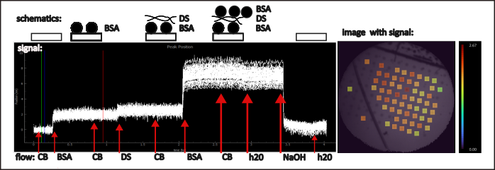
Measurement of adsorption/desorption of a protein monolayer using the integral field camera (IFC) system. A solution of bovine serum albumin (BSA) and dextran sulfate flows over the plasmonic chip, leading to the formation of alternating monolayers on its surface. At the end of the process, the layers are removed by the flow of a basic solution (NaOH). The change in signal caused by adsorption at each individual spot, recorded by the IFC system, is shown in the graph. The curves from each spot exhibit distinct steps, well above the noise level, during protein layer adsorption. The spatial distribution of the signal at each spot after the adsorption of the first BSA layer is shown in the image on the right (signal change from the blue to red line—graph on the left). This distribution is overlaid with the image of the plasmonic chip