
Lensless interferometric scattering microscopy
We developed an ultra-compact optical imaging system for a detection of individual biological species from a liquid solution. The device measures the optical mass of each single object. This can be used in combination with nanoparticle sensors, to count specific molecular species. The device is based on Interferometric Scattering Microscopy (IScat), where a small object adsorbed on an imaging sensor surface forms a small spot with its contrast being proportional to the optical mass. In our concept we combined IScat with Lensless Shadow Imaging (LSI) to remove any optical imaging components. The sensor is formed by the surface of a camera chip being the bottom of the microfluidic chamber. This makes this system extremely robust against vibrations. The number of events is proportional to the concentration. Yet for small concentrations diffusion-limited transport will yield too few collision events with the detection system. We overcome this limit by using magnetic particles. First, molecules (or bacteria) are captured by specific binding to functionalized magnetic particles. Second, the magnetic beads are pulled onto the camera chip via an external magnetic field. The magnetic particles adsorb on the camera chip and its optical mass is determined. The magnetic particles (or molecule-mediated aggregates) with optical mass beyond a bare single particle indicates the presence of the species to detect. The device consists of a low cost CMOS camera, 3D printed fluidic chips and illumination LED. We slightly modify the camera’s hardware/software so that it is applicable for the IScat and magnetic separation. We showed that the device is able to detect single magnetic particles of 100 nm diameter and demonstrated the sensing principle on selective detection of bacteria.
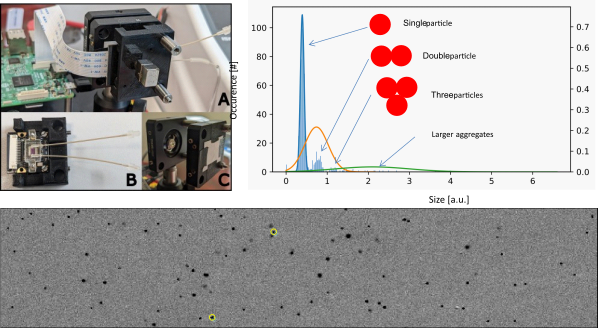
Left - A: a photograph of the core part of the lensless iSCAT system. It consists of a high speed camera with an integrated fluidic chamber (inset B), illumination source (inset C) and permanent magnet, which pulls particles on the surface of the camera chip. Bottom - One time frame of the recorded video with the lensless iSCAT device. These data are processed, so that each spot represent attachment of a single beads at given time and position. Each beads' attachment create a trace of spot with varying contrast. The maximum of the contrast (indicated with yellow circle) is proportional to the 'optical' mass of the beads. Right - a histogram of the contrast value of the particles' attachment events. It is clearly to see the discrete distribution of single particle, particle dimmers and trimers in sample. From this, particle aggregation is quantified.